

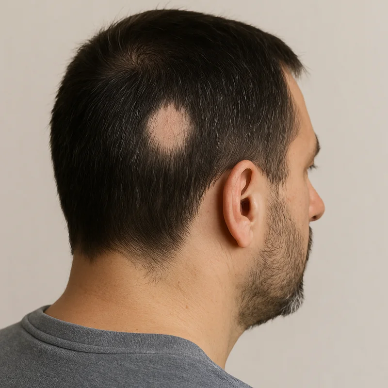
Alopecia areata is the medical term for patchy hair loss – an autoimmune, non-scarring form of hair loss that can affect the scalp, beard, eyebrows and other body hair. Unlike hereditary hair loss, the patches often appear suddenly, and in most cases the follicles are alive but in a dormant state.
This guide covers causes, symptoms, diagnostic findings and treatment options – including PRP treatment and when a hair transplant may be relevant. For a quick overview you can also read our page on alopecia areata treatment under hair loss.
What is alopecia areata?
In alopecia areata the immune system mistakenly attacks hair follicles in the active growth phase (anagen). Normally the follicle has a kind of immune privilege – the immune system largely ignores it. In alopecia areata this protection collapses and cytotoxic T cells gather around the follicles in a pattern sometimes compared to a swarm of bees.
The follicles are forced to stop growing prematurely and enter the resting phase (telogen). The hair shaft loosens and falls out, but the follicle is usually not permanently destroyed.
The condition is divided into three main severity grades:
- Alopecia areata (patchy): patchy hair loss in limited areas
- Alopecia totalis: complete loss of all scalp hair
- Alopecia universalis: total loss of all body hair, including eyebrows, lashes and beard
Causes and risk factors
Alopecia areata causes are multifactorial. Genetic predisposition interacts with immune regulation and external trigger factors.
Genetic factors
Large association studies have identified risk genes in both innate and adaptive immunity. The strongest link is in the MHC region on chromosome 6, where the allele HLA-DQB1*03 occurs in up to 80% of affected individuals in some populations. The disease is also linked to other autoimmune conditions such as APECED syndrome and polymorphisms in immune-regulatory genes.
Stress and environmental factors
Alopecia areata and stress is a well-known association. Under psychological or physical strain the skin’s neuroendocrine system can release neuropeptides that trigger or amplify inflammation around the follicles. This explains why flares often follow major life events – but stress alone rarely causes the disease without underlying genetic vulnerability.
Comorbidities
| Association | Prevalence | Clinical relevance |
|---|---|---|
| Atopic conditions (asthma, eczema, allergic rhinitis) | approx. 60.7% | Strong co-variation – ask about allergy and eczema |
| Vitiligo | approx. 4.1% | Shared autoimmune aetiology |
| Diabetes | approx. 3.2% | Systemic work-up may be warranted |
| Thyroid disease | approx. 2.3% | Thyroid tests may be considered if suspected |
Symptoms and diagnostic findings
Alopecia areata symptoms usually begin as sudden, well-defined round or oval patches of hair loss. The underlying skin is typically smooth, soft and normally coloured, but may show mild redness or itching in the active phase.
Exclamation mark hairs
Exclamation mark hairs are the most specific clinical finding in active alopecia areata. The hairs are broken, usually 2–4 mm long, and taper proximally (towards the scalp) while being thicker distally. They are most visible at the edge of the patch and confirm active disease.
Trichoscopic findings
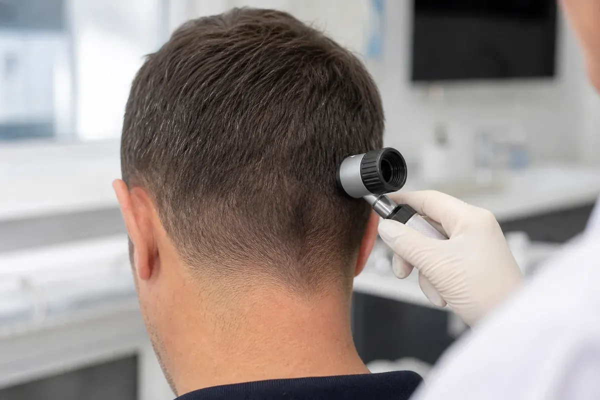
Dermoscopy of the affected area often shows:
- Yellow dots – sebum and keratin debris in dilated follicle openings
- Black dots – pigmented hair fragments broken deep in the follicle
- Poliosis – early grey hair in the regrowth phase
Nail changes
The systemic nature of the disease is sometimes reflected in the nail matrix. Fine pitting, longitudinal ridging and trachyonychia (opaque, rough nail surface) occur in 10.5–17% and are associated with more severe, prolonged courses.
Epidemiology – how common is alopecia areata?
| Parameter | Value | Observation |
|---|---|---|
| Incidence (UK cohort) | 0.26 per 1,000 person-years | Based on 4.16 million individuals |
| Prevalence (adults) | 0.58% | Point prevalence in primary care registers |
| Age at onset | Peak 25–29 years | 85.5% develop before age 40 |
| Ethnic variation | IRR 3.32 (Asian) | Significantly higher incidence in non-white populations |
| Nail involvement | 10.5–17% | Linked to more severe course |
The condition affects both men and women and occurs at all ages, though onset is most common in young adults. In children the course can be more unpredictable.
Psychosocial impact
Although alopecia areata is physiologically benign without direct organ damage, it carries a high psychosocial burden. Hair plays a central role in identity and self-image, and sudden loss can trigger depression and anxiety.
Clinical studies show prevalence of clinical depression at 66.7% and anxiety at 73.3% in patients with alopecia areata. Younger patients (under 30) report high stigmatisation and reduced quality of life. The unpredictable nature of the disease – alternating spontaneous regrowth and sudden relapses – creates constant daily anxiety that in many cases exceeds that seen in potentially life-threatening illnesses.
Alopecia areata in the beard (alopecia barbae)
When the autoimmune process is localised to the beard area it is called alopecia areata beard or alopecia areata barbae. It affects men only and begins as one or more completely bald, round zones in the beard.

For many men the beard is an important marker of masculinity and personal style. Because the face is constantly visible and harder to conceal than the scalp, alopecia areata beard treatment can feel especially urgent. Many shave daily to minimise the visual contrast – which itself can feel like an involuntary loss of identity.
Read more about beard transplantation if you have permanent bald zones after stable disease.
Difference from hereditary hair loss – important differential diagnosis
One of the most important tasks is to distinguish alopecia areata from androgenetic alopecia (hereditary hair loss). Treatment strategies differ fundamentally, and misdiagnosis can lead to ineffective or potentially harmful treatment.
| Factor | Alopecia areata | Androgenetic alopecia |
|---|---|---|
| Pathophysiology | T-cell-mediated autoimmune inflammation | Androgen sensitivity and DHT exposure |
| Time course | Acute, sudden onset | Slowly progressive over years |
| Clinical pattern | Sharply defined round bald patches | Symmetric thinning, receding hairline |
| Distribution | Scalp, beard, eyebrows, body | Strictly androgen-sensitive scalp area |
| Fate of follicle | Intact but dormant | Gradual miniaturisation and atrophy |
| Nails | Pitting may occur | No effect |
| Primary treatment | Corticosteroids, immunotherapy, PRP | Finasteride, hair transplant |
Treatment of alopecia areata
Alopecia areata treatment aims to dampen autoimmune inflammation and stimulate resting follicles to re-enter the growth phase. The choice depends on extent, activity and the patient’s clinical profile.
| Treatment | Indication | Mechanism | Documented effect |
|---|---|---|---|
| Intralesional corticosteroid injections | Limited active patchy alopecia (<50% area) | Local suppression of T-cell attack via triamcinolone acetonide | >50% regrowth in 82.1% after 3 months |
| Contact immunotherapy (SADBE/DPCP) | Extensive, chronic or treatment-resistant alopecia | Controlled allergic dermatitis that redirects immune attack | >50% regrowth in 87.5% after 6 months |
| Systemic methotrexate | Severe, treatment-resistant alopecia | Systemic immunosuppression | Moderate to good effect, high relapse risk on dose reduction |
| PRP | Patchy alopecia, stimulation of weakened follicles | Autologous growth factors stimulate angiogenesis and cell growth | Biologically activates resting follicles |
PRP treatment for alopecia areata
PRP (platelet-rich plasma) is a modern, biological, non-surgical treatment that has shown good results for waking inactive hair follicles. The treatment is based on the patient’s own blood and contains no synthetic additives, minimising the risk of allergic reactions.

Scientific mechanism
In PRP treatment the patient’s blood is centrifuged in a closed, sterile system. Plasma with high platelet concentration (3–5 times higher than normal whole blood) is injected into the affected area. Platelets release biologically active growth factors directly into the follicle microenvironment:
- PDGF (Platelet-Derived Growth Factor)
- VEGF (Vascular Endothelial Growth Factor)
- EGF (Epidermal Growth Factor)
- TGF-β (Transforming Growth Factor-beta)
These growth factors stimulate microcirculation, improve perifollicular oxygenation and induce cell division in follicle stem cells – which can push the follicle out of telogen and re-initiate anagen.
Clinical protocol at Akacia Medical
For patchy hair loss the medical team at Akacia Medical initially recommends a course of three treatments a few weeks apart. After the initial phase results are evaluated carefully:
- Signs of reduced hair loss are often noted after 3–4 weeks
- Visible regrowth may be observed after 3–6 months
To ensure a high concentration of active growth factors, Akacia Medical uses scientifically validated premium systems from Arthrex and RegenLab. Treatment is performed under medical responsibility and strict sterile routines at the clinic in Alvik, Stockholm.
Read more about PRP treatment, prices and hair loss treatments. If you want to understand normal growth rates and when new growth usually becomes visible, read our guide to how fast hair grows.
Hair transplant (FUE) for alopecia areata
A hair transplant with the FUE method is the leading surgical method for permanent hair growth in areas with lasting thinning. In hereditary hair loss individual follicular units are extracted from a stable donor area on the back of the head and transplanted to sparse zones.
For alopecia areata, however, the autoimmune pathology places extremely strict requirements on patient selection:
| Clinical status | Suitability for FUE | Rationale |
|---|---|---|
| Active alopecia areata (ongoing flare) | Absolutely unsuitable | Autoimmune attack rejects newly transplanted follicles – permanent loss of donor hair |
| Unstable or fluctuating disease | Unsuitable | New patches elsewhere indicate systemic instability |
| Alopecia totalis / universalis | Absolutely unsuitable | No healthy donor area exists |
| Long-term stable, burnt-out alopecia | May be considered | Remission for at least 2–3 years and fibrotic, burnt-out patches |
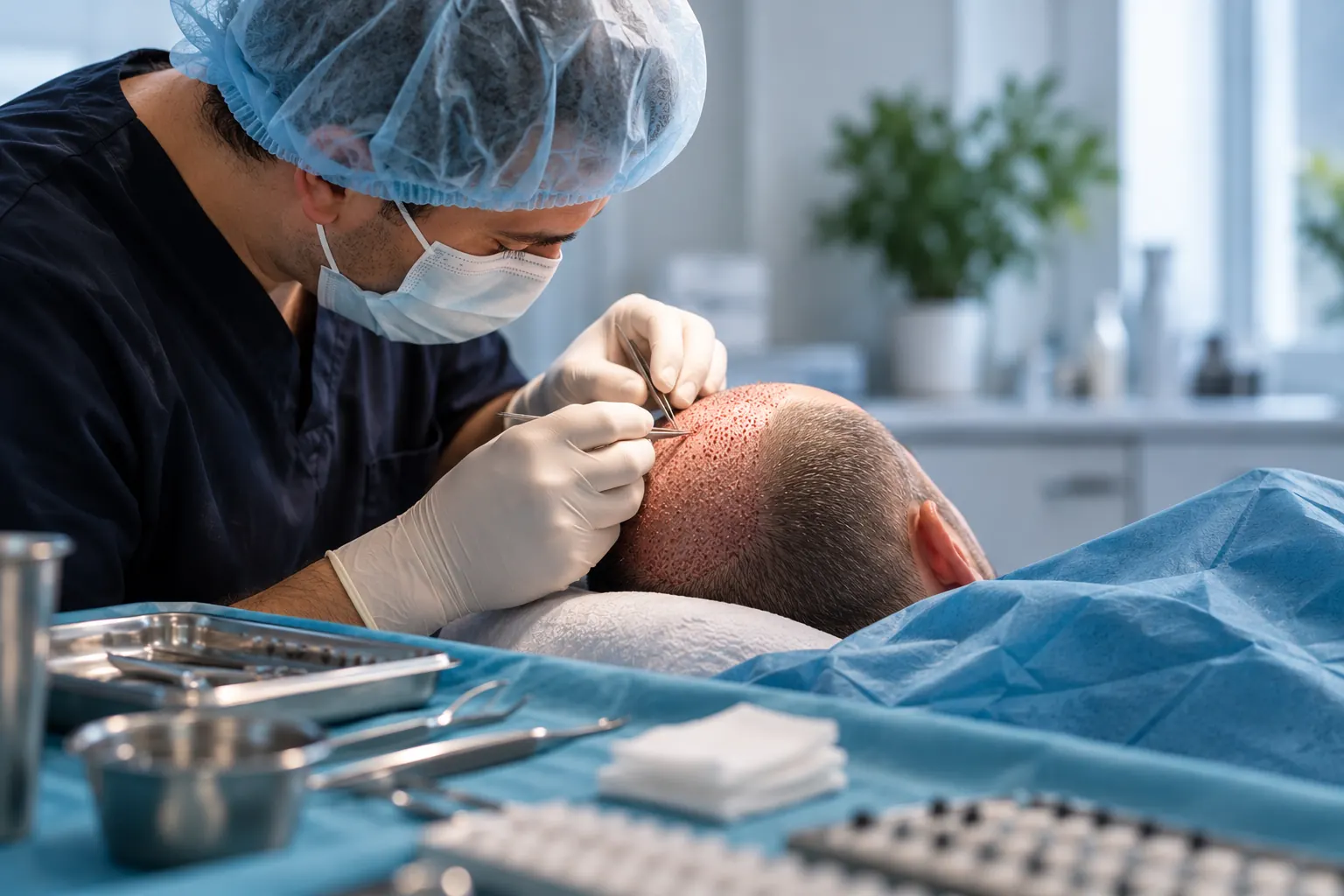
When a patient meets the criteria for a stable, burnt-out phase, FUE hair transplantation may be considered in selected cases after careful medical assessment. The procedure is performed under local anaesthesia and is essentially painless. Akacia Medical places each graft manually with precision regarding angle, depth and direction – with experience since 2011, licensed doctors and ISHRS membership.
Summary and clinical guidance
Alopecia areata and its beard variant, alopecia barbae, are complex autoimmune conditions that require medical expertise and realistic expectations. Unlike hereditary hair loss, follicles are in most cases alive but dormant – which means non-surgical stimulation techniques should be prioritised in the initial phase.
Recommended action plan
- Diagnosis: get a professional assessment to distinguish alopecia areata from other forms of hair loss. Start with our page on alopecia areata treatment.
- Stimulation: consider PRP treatment if you want to stimulate resting follicles biologically under controlled conditions.
- Permanent restoration: in long-term stable, burnt-out disease, hair transplantation with FUE may be relevant – but never during active disease.
Once the condition is stable, a hair transplant may be an option. We provide hair transplant Stockholm care and hair transplant in Sweden.
Next steps
- Book a free consultation – in Stockholm or Gothenburg, or online or by phone
- See prices and packages for PRP and hair transplantation
- Read about hair loss treatments and which options suit your situation
This text is for informational purposes only. For medical advice or diagnosis, consult a qualified professional.
Frequently asked questions
Tags
Related information
Explore more about our treatments and services

Firo Esmer
CEO, Akacia Medical
Firo Esmer is CEO and founder of Akacia Medical and writes about hair transplantation, hair loss and treatment planning based on the clinic's experience and patient journey.
References
- 1.Harries M et al. Epidemiology of alopecia areata: a population-based cohort study in UK primary care. Br J Dermatol. 2022.
- 2.Gilhar A et al. Lymphocytes, neuropeptides, and genes involved in alopecia areata. J Clin Invest. 2007.
- 3.Ihm CW, Han JH. Diagnostic value of exclamation mark hairs. Dermatology. 1993.
- 4.Tosti A, Bellavista S, Iorizzo M. Alopecia areata: a long term follow-up study of 191 patients. J Am Acad Dermatol. 2006.
- 5.Phan K, Ramachandran V, Sebaratnam DF. Methotrexate for alopecia areata: A systematic review and meta-analysis. J Am Acad Dermatol. 2019.
- 6.Ghaffari J et al. Association among Thyroid Dysfunction, Asthma, Allergic Rhinitis and Eczema in Children with Alopecia Areata. Open Access Macedonian J Med Sci. 2017.
- 7.Tan E, Tay YK, Goh CL, Chin G. The pattern and profile of alopecia areata in Singapore. Int J Dermatol. 2002.
Related articles
September 10, 2026
Does a hair transplant hurt?
Does a hair transplant hurt? How local anaesthesia works, what you feel during the procedure and how tenderness is handled in the first days.
September 3, 2026
Hair transplant scars – FUE, FUT and healing
How visible are scars after a hair transplant? The difference between FUE and FUT, healing in the donor area, and how older scars can be hidden.
August 14, 2026
How many hair transplants can you have?
How many hair transplants can you have? Donor area, healing and planning set the limit. Learn about treatments, timing and consultation.
Book a free consultation
We will review your circumstances and recommend the right next step for you.